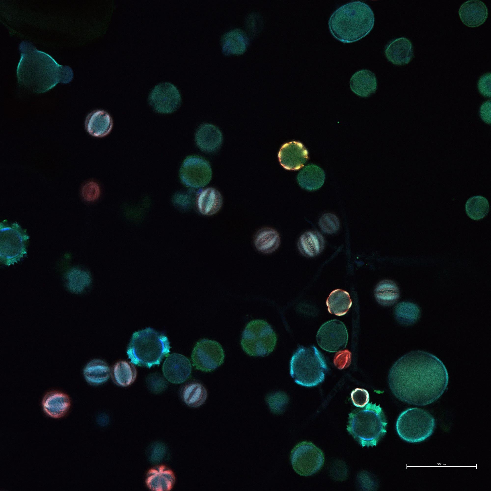
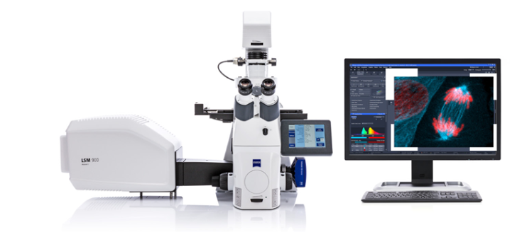
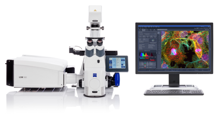
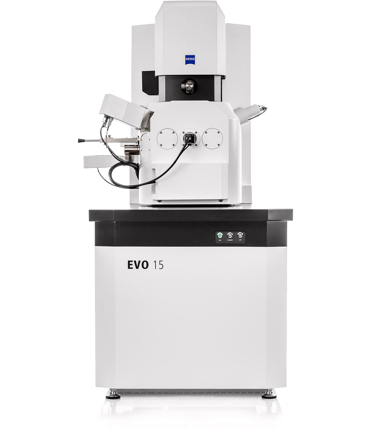
The Imaging Unit of the CFH (CFH-IMG) provides research services in the fields of light microscopy, confocal laser scanning microscopy, scanning electron microscopy, Raman microscopy, atomic force microscopy and certain correlative combinations thereof, as well as the development of new analytical methods in the above fields, primarily for members of the University of Hohenheim, but also for external users. The CFH-IMG operates in two modes:
Advanced confocal laser scanning microscope with motorized inverted stand Axio Observer
7.
The system is equipped with four lasers (405, 488, 561, 640 nm) and can be used for epifluorescence and
brightfield microscopy.
A 3-channel GASP spectral detector captures the emission light and enables
qualitative and quantitative analyses. Alternatively, the system is also equipped with a color
camera.
Different deconvolution principles (Airyscan 2, LSM Plus) facilitate a high signal-to-noise ratio. The
detection speed can be increased by multiplexing.
Resolution ranges from 488 nm to 90/120 nm, with common deconvolution and excitation wavelength.
Extensive hardware, accessories, upgrades (e.g., incubation chamber, wide range of objectives) and
software equipment are available for life cell
imaging, as well as for correlative microscopy with SEM/STEM.
Powerful workstations with
state-of-the-art analysis software (ZEN Module, arivis Vision4D) are available to users.
State-of-the-art confocal laser scanning microscope with Axio Observer 7 motorized inverted stand and
automatic specimen detection.
The system is equipped with five lasers (405, 488, 561, 594,633 nm) and a tunable pulsed multiphoton
laser (690-1040nm), and can be used for epifluorescence as well as for reflected light microscopy.
Two GASP-NDD detectors and a 34 channel spectral detector allow single photon counting. Overlapping
fluorochromes and autofluorescence can be separated online in one scan. Both qualitative and
quantitative analyses are possible.
Different deconvolution principles (Airyscan 2, LSM Plus) in confocal as well as in
multiphoton mode facilitate a high signal-to-noise ratio. The detection speed can be increased by
multiplexing (SR-8Y).
The resolution capability is 90/120 nm when using joint deconvolution and an
excitation wavelength of 488 nm.
Extensive hardware, accessories, upgrades (e.g., incubation chamber, wide
range of objectives) and software equipment are available for life cell imaging, as well as for
correlative
microscopy with SEM/STEM.
Powerful workstations with state-of-the-art analysis software (ZEN Module, arivis
Vision4D) are available to users.
High-resolution scanning electron microscope (SEM) with LaB6 cathode, camera in the
sample chamber
and three-lens electron optics with beam control.
Spatial resolution in high vacuum and use of
secondary electron detector of 3.0 nm at 30 kV, and 8.0 nm at 3 kV. Secondary electron detection is
possible in high and low vacuum operation.
Magnification is continuously adjustable in the range of
5 - 1,000,000 x, accelerating voltage ranges 0.2 - 30kV.
Thin tissue sections can be examined using
a STEM (Scanning Transmission Electron Microscopy) detector.
The sample chamber has an inner
diameter of 365 mm and a height of 275 mm and is loaded via the sample chamber door. Samples are
loaded into the instrument on a 5-axis motorized Cartesian stage (X,Y,Z = 125 x 125 x 50 mm), 360°
rotation and tilting (-10° to 90°) is possible in three spatial directions.
An EDX detector (100 mm²
detector area) allows elemental analysis on a microscopic scale.
In addition, full equipment for
correlative microscopy with a light microscope is available.
Correlative Raman-AFM system to combine chemical imagine with high-resolution surface
analysis.
Confocal Raman microscopy with various recoding modes, e.g. planar scans in x-, and y-dimensions,
in-depth scans or z-stacks and 2D and 3D Raman Imaging with Raman spectra recording at each
pixel.
The system contains two different laser sources (green 532nm, and near infrared 785nm), coupled with
a spectrometer of various gratings for Raman measurements.
The objective turret holds up to five different objectives. Six different objectives are available
ranging from 10x to 100x with one dedicated for AFM measurements.
Both Raman and AFM functionality can be operated and easily switched within the WiTec project SIX
software. The software package contains additional features like WITec Particle Scout for particle
search and classification and WITec TrueMatch database management for particle identification with a
S.T.Japan dataset of biochemicals, consisting of more than 4,000 entries.