Examples
Nutrient density analysis in regeneratively farmed produces
In this EIT Food project, we analyzed the principal macronutrients (proteins, carbohydrates and lipids) and micronutrients (vitamins and minerals) to determine nutrient density present in produces from regenerative agriculture where there has been a special focus on soil health and biodiversity. By comparing the nutrient density of regeneratively farmed produce to the same produce from conventional agriculture, the project investigated the a correlation between healthy soils and healthy produces. EIT Food is Europe's leading food innovation initiative working to make the food system more sustainable, healthy and trusted.
TerrAquat
Ongoing care and monitoring of agricultural soils are a prerequisite for building a sustainable and economically viable agri-food system. Increasing challenges of industrialized and extensive production systems and scientific findings also lead to a growing need for innovative solutions to carefully assess the health of soils, plants and their interconnected ecosystems. "TerrAquat" has developed self-integrating accumulators (SIA) that enable continuous monitoring of soil and water quality. The collaboration with the CFH focuses on assessing nutrient leaching, e.g. nitrates and ammonia. SIAs are permanently installed in the soil to assess their filtering ability and preferential flow of leachate. They are harvested twice a year, subjected to extraction, and soluble nitrogen compounds are subsequently quantified using photometric methods.
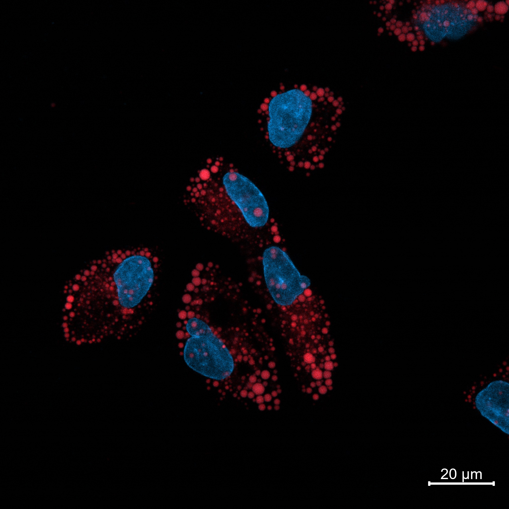
Lipid Droplets in HepG2 Cells
This microscopy image showcases HepG2 cells incubated with fatty acids and stained with DAPI and Nile Red.
The red fluorescence highlights lipid droplets stained by Nile Red, while the blue fluorescence represents cell nuclei stained with DAPI.
This dual staining method provides clear visualization of lipid storage and cellular structures, offering valuable insights into lipid metabolism and cell biology.
.jpg)
Pollen Analysis
Pollen analysis belongs to food analysis and consists of the examination of pollen
in honey. Pollen is ingested by bees with nectar when they visit flowers. Pollen can be
identified under the light microscope and assigned to the plants of origin. Confocal light
microscopy makes use of the fact that pollen has a pronounced intrinsic fluorescence and emits
fluorescent light of different wavelengths after excitation, even without additional
staining.
The pollen analysis is thereby an indirect proof, how high the nectar portion of a certain plant
is at the honey and is used with the examination of the sort indication. The honey thus provides
a reflection of the plants visited by the bee during nectar collection. The plant origin of the
honey and its plant spectrum can thus also provide information about its geographical origin.
Animal alternative products
Sustainability, a plant-based diet and regenerative agriculture represent some of the top food trends for 2023. Consumers engage more and more with food and want to relate with its origin and nutritiousness, as well as its effect on animal welfare and the environment. This increases pressure on food companies, which must constantly present innovative products to meet these expectations and maintain competitiveness. In particular, the area of plant-based alternatives presents a major challenge, as plant-based ingredients differ greatly in composition, usability and handling from animal protein sources such as milk and meat. This leads to changes in texture and mouthfeel when comparing, for example, traditional fresh cheese and fresh cheese analogs. The use of confocal light microscopy enables structural elucidation of these products, as protein, fat and carbohydrate compounds can be individually stained and visualized to understand and optimize the emulsion and bulk properties of plant-based analogs.
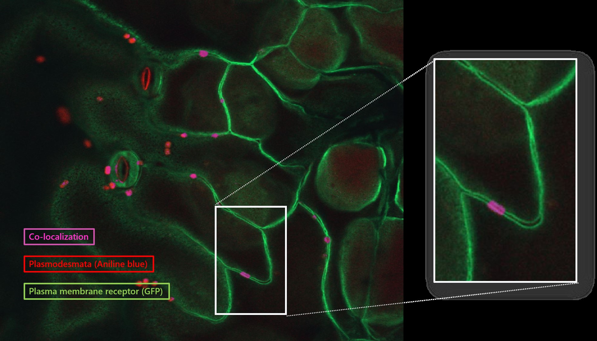
Co-localization of a Receptor Like Kinase and Plasmodesmata
Plasma membrane Receptor-like kinases perceive environmental stimulus and transient
them as a signal downstream to the cell in order to regulate plant growth or perform the stress
defense
Plasmodesmata is the most special structure in plant, acts as the gates in cell wall mediating
dynamic cell-to-cell communication. as an cell-to-cell gate between different cells.
We found certain plasma membrane receptor-like kinases could localize plasmodesmata.
How can we observe the co-localization?
Anilin blue stains Plasmodesmata; the receptor like kinase-GFP localizes at plasma membrane